
2026/11/14 09:00-10:30 Room 401
- Taiwan-Korea Stroke Research Forum
- Time
- Topic
- Speaker
- Moderator
- 09:40-10:00
- Thrombus Ultrastructure: From Electron Microscopy to Imaging and Clinical Implications
- SPEAKER:
Hyungjong Park
Korea,South (南韓)
- MODERATOR:
Hyo Suk Nam
Korea,South (南韓)
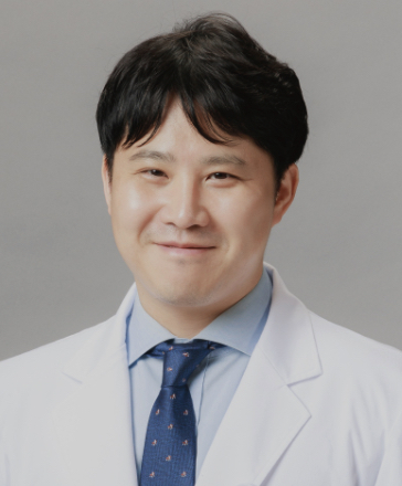
- Hyungjong Park
- MD, MSc
-
Associate Professor, Department of Neurology, School of Medicine, Kyungpook National University
Department of Neurology, Kyungpook National University Hospital.,
Lecture Abstract:
Histological analysis tells us what a retrieved thrombus is made of. Electron microscopy tells us how it is built. Scanning electron microscopy (SEM) resolves surface architecture at nanometer scale, showing the deformation of red blood cells from biconcave discs into tightly packed polyhedra, fibrin appearing as radiating bundles or amorphous sponge, platelet aggregates, neutrophil extracellular traps, and the empty space between these components. Transmission electron microscopy adds the intracellular and membrane-level detail that surface imaging cannot provide. Since the 2017 international consensus statement recommended EM as a complementary modality in clot research, these techniques have moved from descriptive morphology toward mechanistically meaningful measurement. This presentation will outline three connections. First, ultrastructure and biology: polyhedral erythrocyte deformation is a signature of platelet-driven clot contraction, and a compact outer shell is one explanation for fibrinolysis resistance. Second, ultrastructure and procedure: clot compactness, elasticity, and surface texture influence adhesion to stent retrievers and to aspiration catheters, and therefore relate to pass count and procedural duration. Third, ultrastructure and imaging: the same architecture that determines mechanical behavior also determines contrast penetration, making clot perviousness on CT a candidate non-invasive surrogate for what EM shows only after retrieval. The central limitation is practical. EM is destructive, slow, sampling-dependent, and manually quantified, which confines it to retrospective research rather than periprocedural decision-making. I will close by introducing our ongoing work applying deep learning to EM image analysis, using automated segmentation and quantification of clot components, with the aim of making ultrastructural phenotyping reproducible, scalable, and ultimately linkable to pre-treatment imaging.
Histological analysis tells us what a retrieved thrombus is made of. Electron microscopy tells us how it is built. Scanning electron microscopy (SEM) resolves surface architecture at nanometer scale, showing the deformation of red blood cells from biconcave discs into tightly packed polyhedra, fibrin appearing as radiating bundles or amorphous sponge, platelet aggregates, neutrophil extracellular traps, and the empty space between these components. Transmission electron microscopy adds the intracellular and membrane-level detail that surface imaging cannot provide. Since the 2017 international consensus statement recommended EM as a complementary modality in clot research, these techniques have moved from descriptive morphology toward mechanistically meaningful measurement. This presentation will outline three connections. First, ultrastructure and biology: polyhedral erythrocyte deformation is a signature of platelet-driven clot contraction, and a compact outer shell is one explanation for fibrinolysis resistance. Second, ultrastructure and procedure: clot compactness, elasticity, and surface texture influence adhesion to stent retrievers and to aspiration catheters, and therefore relate to pass count and procedural duration. Third, ultrastructure and imaging: the same architecture that determines mechanical behavior also determines contrast penetration, making clot perviousness on CT a candidate non-invasive surrogate for what EM shows only after retrieval. The central limitation is practical. EM is destructive, slow, sampling-dependent, and manually quantified, which confines it to retrospective research rather than periprocedural decision-making. I will close by introducing our ongoing work applying deep learning to EM image analysis, using automated segmentation and quantification of clot components, with the aim of making ultrastructural phenotyping reproducible, scalable, and ultimately linkable to pre-treatment imaging.