學會會訊

會員登入

帳號

密碼


神經影像學病例報告

作者:陳右緯 (壢新醫院神經內科主任)

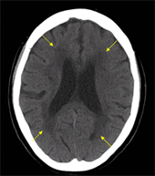
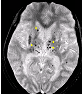
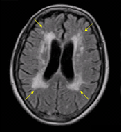
65歲女性廚師,因為前一日突發性的右側偏癱及構音困難住院。過往病史包括一次短暫性黑矇症,以每日Aspirin 100mg治療;高血壓,規則藥物控制;高血脂,以飲食控制。住院時意識清楚,神經學檢查顯示構音困難、右側偏癱含中樞型顏面神經麻痺,NIHSS=5。住院時腦部電腦斷層及核磁共振影像學如下。

圖A:頭部CT:箭頭處為兩側腦室旁白質病變。嚴重程度的計分法有很多種,筆者習慣使用的為van Sweieten 之計分法(1)。本法取經過後角 (Posterior Horn)的脈絡叢、側腦室中心區(Cella Media)及Centrum Semiovale的三個切面來評估,以較嚴重的一邊為準。將大腦分為前後兩區分別評估,分別為0 (無白質病變)、1 (具腦室旁白質病變,但未到達灰白質交界)、2 (白質病變到達灰白質交界)。前後大腦分數相加,總分為0至4分。本案例總分為2分。圖B:MRI (FLAIR-weighted)因將腦室之腦脊髓液呈現為低訊號,較傳統T2-weighted MRI更能顯出白質病變之範圍,以供定量分析比較(2)。圖C與圖D:Gradient Echo (T2*-weighted) MRI:多處箭頭指出低訊號處為腦部微出血。

腦部微出血的分布可能和不同的血管病變有關,於高血壓或粥狀動脈硬化病患常見於基底核、小腦及腦幹(3);而侷限於腦葉者則常見於類澱粉血管病變相關,尤其是大腦後側之頂葉、顳葉及枕葉(4,5)。雖然針對急性腦梗塞患者之靜脈血栓溶解治療可延伸至發病後四個半小時內注射(6),但是已有許多報告指出具有白質病變或腦部微出血病灶之患者施打血栓溶解劑後,腦出血機會有增加的趨勢(7,8)。影像學的資訊是否會影響未來的治療指引,有待更多的臨床研究結果。
 
圖A 圖C
圖B 圖D
 
參考文獻:
  1. van Swieten JC, Hijdra A, Koudstaal PJ, et al. Grading white matter lesions on CT and MRI: a simple scale. J Neurol Neurosurg Psychiatry 1990;53:1080-3.
  2. Chen YW, Gurol ME, Rosand J, et al. Progression of white matter lesions and hemorrhages in cerebral amyloid angiopathy. Neurology 2006;67:83-7.
  3. Vernooij MW, van der Lugt A, Ikram MA, et al. Prevalence and risk factors of cerebral microbleeds: the Rotterdam Scan Study. Neurology 2008;70:1208-14.
  4. Lee SH, Kim SM, Kim N, et al. Cortico-subcortical distribution of microbleeds is different between hypertension and cerebral amyloid angiopathy. J Neurol Sci 2007;258:111-4.
  5. Rosand J, Muzikansky A, Kumar A, et al. Spatial clustering of hemorrhages in probable cerebral amyloid angiopathy. Ann Neurol 2005;58:459-62.
  6. Hacke W, Kaste M, Bluhmki E, et al. Thrombolysis with alteplase 3 to 4.5 hours after acute ischemic stroke. N Engl J Med 2008;359:1317-29.
  7. Neumann-Haefelin T, Hoelig S, Berkefeld J, et al. Leukoaraiosis is a risk factor for symptomatic intracerebral hemorrhage after thrombolysis for acute stroke. Stroke 2006;37:2463-6.
  8. Fiehler J, Albers GW, Boulanger JM, et al. Bleeding risk analysis in stroke imaging before thromboLysis (BRASIL): pooled analysis of T2*-weighted magnetic resonance imaging data from 570 patients. Stroke2007;38:2738-44.

對於此文章如有任何問題或指教,歡迎來信至學會,編輯部後續會將您的意見刊載在下一期會訊!